Respiratory system
Respiratory system
The respiratory system (or ventilatory system) is the biological system of an organism that introduces respiratory gases to the interior and performs gas exchange.
In humans and other mammals, the anatomical features of the respiratory system include airways, lungs, and the respiratory muscles.
Molecules of oxygen and carbon dioxide are passively exchanged, by diffusion, between the gaseous external environment and the blood.
This exchange process occurs in the alveolar region of the lungs.
Lung
The lung is the essential respiration organ in many air-breathing animals, including most tetrapods, a few fish and a few snails.
In mammals and the more complex life forms, the two lungs are located near the backbone on either side of the heart.
Their principal function is to transport oxygen from the atmosphere into the bloodstream, and to release carbon dioxide from the bloodstream into the atmosphere.
This exchange of gases is accomplished in the mosaic of specialized cells that form millions of tiny, exceptionally thin-walled air sacs called alveoli.
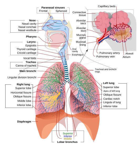
To completely explain the anatomy of the lungs, it is necessary to discuss the passage of air through the mouth to the alveoli.
Once air progresses through the mouth or nose, it travels through the oropharynx, nasopharynx, the larynx, the trachea, and a progressively subdividing system of bronchi and bronchioles until it finally reaches the alveoli where the gas exchange of carbon dioxide and oxygen takes place.
The drawing and expulsion of air (ventilation) is driven by muscular action; in early tetrapods, air was driven into the lungs by the pharyngeal muscles via buccal pumping, whereas in reptiles, birds and mammals a more complicated musculoskeletal system is used.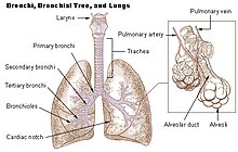
Trachea
In tetrapod anatomy the trachea, or windpipe, is a tube that connects the pharynx or larynx to the lungs, allowing the passage of air.
It is lined with pseudostratified ciliated columnar epithelium cells with goblet cells that produce mucus. This mucus lines the cells of the trachea to trap inhaled foreign particles that the cilia then waft upward toward the larynx and then the pharynx where it can be either swallowed into the stomach or expelled as phlegm.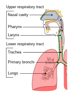
Diaphragm
In the anatomy of mammals, the thoracic diaphragm, or simply the diaphragm , is a sheet of internal skeletal muscle that extends across the bottom of the rib cage.
The diaphragm separates the thoracic cavity (heart, lungs & ribs) from the abdominal cavity and performs an important function in respiration: as the diaphragm contracts, the volume of the thoracic cavity increases and air is drawn into the lungs.

Project's hierarchical decomposition
To create the project in 3D on the respiratory system, i started by building the lungs initially.
Human lungs are located in two cavities on either side of the heart.
Both are separated into lobes by fissures, with three lobes on the right and two on the left.
The lobes are further divided into segments and then into lobules, hexagonal divisions of the lungs that are the smallest subdivision visible to the naked eye.
Each lobe is surrounded by a pleural cavity, which consists of two pleurae. The parietal pleura lies against the rib cage, and the visceral pleura lies on the surface of the lungs.
In between the pleura is pleural fluid. The pleural cavity helps to lubricate the lungs, as well as providing surface tension to keep the lung surface in contact with the rib cage.
The Lung
The lungs are divided mainly by multiple lobes, so I decided to decompose each lung into two parts: an upper and a lower representatives of the division created by the different lobes.
Both the upper and the lower part have a shape not square but curvilinear.
For this reason for their realization has been necessary to use BEZIER sectioning each part in more curves, always using this function, and approximating the same so as to create a surface.
The diversity in the two parts is that the final section of the lower part, was created using HERMITE as it is a reentrant surface as it has a cavity in which rely other organs of the digestive system.
Once created, the two parties, to avoid marking the separation between the lobes too, was chosen to approximate the last curve of the top with the bottom starting from the first curve of the latter. Procedure performed again using BEZIER.
We also know that the two lungs are similar and symmetrical to each other for this, after creating the first lung, the second has been created by simple rotation and translation of the first.
The first image represents the 3d lugs in Plasm.js.


The Trachea,bronchi and bronchioles
The trachea has an inner diameter of about 25.4 millimetres (1.00 in) and a length of about 10 to 16 centimetres (3.9 to 6.3 in) and it's shape is almost cylindrical, generated by CYL_SURFACE.
There are about fifteen to twenty incomplete C-shaped cartilaginous rings that reinforce the anterior and lateral sides of the trachea to protect and maintain the airway, leaving a membranous wall (pars membranacea) dorsally without cartilage.
The trachealis muscle connects the ends of the incomplete rings and contracts during coughing, reducing the size of the lumen of the trachea to increase the air flow rate.
These rings have been created through nl'estrusione of various discs of a diameter slightly larger than the trachea.
The trachea and bifurcates into the primary bronchi at the vertebral level of thoracic vertebra, or up to two vertebrae lower or higher, depending on breathing.
Primary bronchi were created using a function that takes as input height and thickness, which rotates symmetrically a cylindrical surface having the same extruded surface from small disks as in the case of the trachea.
The segmental bronchi divide into many primary bronchioles which divide into terminal bronchioles, each of which then gives rise to several respiratory bronchioles, which go on to divide into two alveolar ducts.
The bronchioles were created with a similar function to that used for the bifurcation of the trachea in the primary bronchi, with the difference that, in addition to the bifurcation, is also represented the main section.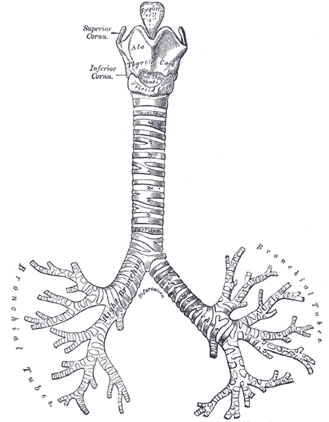 This image represents trachea,bronchi and bronchioles in Plasm.js.

The Diaphragm
The diaphragm functions in breathing.
During inhalation, the diaphragm contracts, thus enlarging the volume of the thoracic cavity (the external intercostal muscles also participate in this enlargement).
This reduces intra-thoracic pressure: In other words, enlarging the cavity creates suction that draws air into the lung, or lungs.
The diaphragm is a dome-shaped musculofibrous septum that separates the thoracic from the abdominal cavity, its convex upper surface forming the floor of the former, and its concave under surface forming the roof of the latter.
Its peripheral part consists of muscular fibers that take origin from the circumference of the inferior thoracic aperture and converge to be inserted into a central tendon.
For its realization, because of its curvilinear shape, has been thought to use both HERMITE that BEZIER.
Diaphragm in Plasm.js:

Final result
This image represents the final project in 3D respiratory launched on Google Chrome using the platform framework Plasm.js.

References
Wikipedia;
All the images used for the realization of this project can be found at this link.
The Human Body, An Illustrated Guide to Every Part of The Human Body and How it Works;
My cvdlab-cg Home;
My final-project.